
This is a proof of concept that
allows imaging several millimeters deep into tissue. It will certainly end up first been used with
cardiovascular situations.
It works by activating bonds in
fat and that provides the structural detail.
Specific molecules can be mapped.
The obvious target is plaques and
that is what will be gone after. Yet
this must also be useful in investigating small tumors.
Our imaging technology continues
to improve and will continue to do so.
New microscope peers deep into tissue
Aug 5, 2011 1 comment
Researchers in the US
In recent years, scientists have developed microscopy techniques that
can locate specific molecules in a biological sample without the need to label
those molecules. Although techniques such as stimulated Raman scattering and
coherent anti-Stokes Raman scattering have revolutionized biological imaging,
their use is limited by their relatively small penetration depth.
Now, a team led by Ji-Xin Cheng at Purdue University
Picking up vibrations
The technique involves firing a laser pulse at a sample to excite a
specific vibrational mode associated with the carbon–hydrogen bonds that abound
in body fat. The wavelength of the pulse is chosen so that absorption by
blood and surrounding tissue is minimal. The laser pulses cause the fat
molecules to heat and expand locally, thus generating pressure waves at
ultrasound frequencies that are detected by a transducer.
By scanning the laser over the sample in 2D and measuring the arrival
time and intensity of the ultrasound at a number of different locations, the
team is able to create a 3D image giving the location of fat in the sample.
"Targeting specific chemical bonds is expected to open a
completely new direction for the field," says Cheng. "Measuring the
time delay between the laser and the ultrasound waves gives a precise distance,
which enables you to image layers of tissue and create 3D pictures using just
one scan."
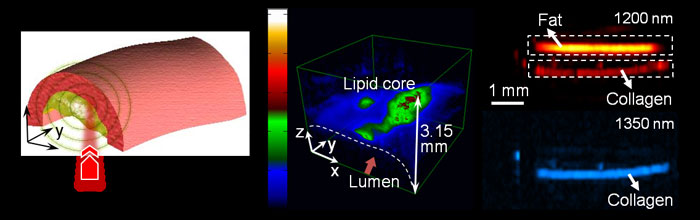
To demonstrate the potential of 3D VPA imaging, carotid arteries were
removed from pigs with profound atherosclerosis. The team detected a strong
VPA signal from fat molecules located 1.5 mm below the illuminated surface
of the sample, allowing the identification of different levels of fat
accumulation. The VPA technique clearly distinguished a number of different
fatty deposits in the arteries. This is important in the study and diagnosis of
cardiovascular diseases because fat combines with other substances to form
artery-clogging plaque. The researchers also used VPA microscopy to map the
distribution of fats in fruit-fly larvae.
Next step is miniaturization
The Purdue group is now looking to miniaturize its system and develop a
catheter-based imaging device. "We are hoping to build an endoscope to put
into blood vessels," says Cheng. "This would enable us to see the
exact nature of plaque formation in the walls of arteries and to better
quantify and diagnose cardiovascular disease."
Team member Han-Wei Wang adds that the spatial resolution of the VPA
system is suitable for such future work. "The lateral resolution is very
flexible from the order of a micrometre to tens of micrometres," he says.
"The resolution is an improvement compared with current clinical imaging
methods such as intravascular ultrasound. Our spatial resolution will be enough
for atherosclerotic applications, and will be a great option as a complementary
imaging modality."
Although the first area of interest for the Cheng group is
cardiovascular disease, in the future the method might also be used to detect
fat molecules in muscles to diagnose diabetes or other lipid-related disorders,
including neurological conditions and brain trauma. The technique can also
image protein fibrils, making it useful when studying collagen's role in scar
formation.
The work is described in Physical Review Letters.
About the author
Jacqueline Hewett is a freelance science and technology journalist
based in Bristol, UK, and Hamish
Johnston is editor ofphysicsworld.com
No comments:
Post a Comment